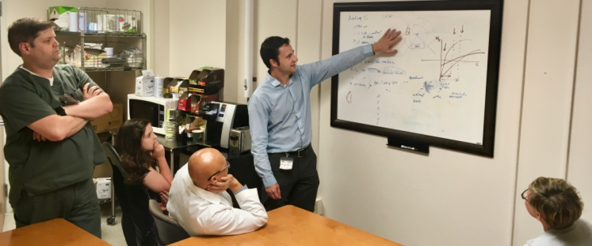
Research is one of the three major programs within the DCMRC and is designed to be fully integrated with the Patient Care and Education programs. The Research program itself involves multiple approaches: from basic development of new technology such as novel pulse sequences, to in-vivo testing in physiology models, to translational studies in humans validating clinical utility, to multi-center clinical trials. Funding for the Center is provided by grants from the National Institutes of Health, American Heart Association, and Industry.
An important aspect of research activities at the DCMRC is the development of new MRI pulse sequences and imaging protocols. The DCMRC has dedicated workstations for modifying MRI scanner software at its lowest level, as well as individuals from academia and industry whose primary responsibility is sequence and protocol development. Candidate pulse sequences and protocols are first evaluated at the Duke South facility, and those whose clinical and/or research potential can be demonstrated are added to the library of active scan protocols.
The DCMRC is also active in multi-center clinical trials. Examples of DCMRC run trials include the first multi-center trial designed to evaluate an imaging approach for the detection of myocardial infarction, and the APEX-AMI CMR sub study. The DCMRC has or is currently providing MR core lab services for several industry sponsored multi-center clinical trials (including from Proctor and Gamble, Boston Scientific, Mallinckrodt, TargeGen, and Velocimed) and NIH sponsored multi-center studies (including REVEAL and RELAX).
The DCMRC administrative and research space is located in the Duke Clinic and hosts MR core laboratory services. These services include consultation with the PI for developing MR imaging protocols designed to evaluate the specified study endpoint(s), site selection, site initiation, quality review of scans as they are received, rapid feedback to sites regarding the quality of the images and adherence to the imaging protocol, qualitative and quantitative assessment of specified study endpoints, and collaboration with the PI regarding the results of the study relative to the cardiovascular MR endpoints. The workspace is equipped with imaging analysis workstations, desktop computers, and a high-speed network for post-doctoral research associates, clinical fellows, and other staff participating in research. A dedicated web-based PACS that allows a bird's eye view of all images received from all sites is utilized for core lab services.
Please contact Michele Parker (michele.parker@duke.edu, 919-668-1671) if you have a project or trial that requires the services of a central CMR core laboratory.
Recent Notable Publications
Kim, HW, Rehwald, WG, Jenista, ER, Wendell, DC, Filev, P, van Assche, L, Jensen, CJ, Parker, MA, Chen, EL, Crowley, ALC, Klem, I, Judd, RM, Kim, and RJ. "Dark-Blood Delayed Enhancement Cardiac Magnetic Resonance of Myocardial Infarction." JACC Cardiovascular Imaging, December 2017, (epub ahead of print) Full Text
Kim, HW, and Kim, RJ. "Prognostic Value of Myocardial Damage in Patients With Sarcoidosis: Is Cardiac Magnetic Resonance What We Need?." Circulation. Cardiovascular Imaging 9, no. 9 (September 2016): e005518- Full Text
Chang, S-A, and Kim, RJ. "The Use of Cardiac Magnetic Resonance in Patients with Suspected Coronary Artery Disease: A Clinical Practice Perspective." Journal of Cardiovascular Ultrasound 24, no. 2 (June 22, 2016): 96-103. (Review) Full Text
Jenista, ER, Rehwald, WG, Chaptini, NH, Kim, HW, Parker, MA, Wendell, DC, Chen, E-L, and Kim, RJ. "Suppression of ghost artifacts arising from long T1 species in segmented inversion-recovery imaging." Magnetic resonance in medicine (November 20, 2016). Full Text
Kim, HW, Van Assche, L, Jennings, RB, Wince, WB, Jensen, CJ, Rehwald, WG, Wendell, DC, Bhatti, L, Spatz, DM, Parker, MA, Jenista, ER, Klem, I, Crowley, ALC, Chen, E-L, Judd, RM, and Kim, RJ. "Relationship of T2-Weighted MRI Myocardial Hyperintensity and the Ischemic Area-At-Risk." Circulation research 117, no. 3 (July 2015): 254-265. Full Text
Smulders, MW, Bekkers, SCAM, Kim, HW, Van Assche, LMR, Parker, MA, and Kim, RJ. "Performance of CMR Methods for Differentiating Acute From Chronic MI." JACC. Cardiovascular imaging 8, no. 6 (June 2015): 669-679. Full Text
Smulders, MW, Bekkers, SCAM, Kim, HW, Van Assche, LMR, Parker, MA, and Kim, RJ. "Performance of CMR methods for differentiating acute from chronic MI." JACC: Cardiovascular Imaging 8, no. 6 (January 1, 2015): 669-679. Full Text
Klem, I, Heiberg, E, van Assche, L, Wagner, G, Parker, M, Kim, HW, Grizzard, JD, Arheden, H, and Kim, R. "Sources of variability in quantification of CMR infarct size and their impact on sample size calculations - reproducibility among three core laboratories." Journal of Cardiovascular Magnetic Resonance 17, no. Suppl 1 (2015): P84-P84. Full Text
Heitner, JF, Klem, I, Rasheed, D, Chandra, A, Kim, HW, Van Assche, LMR, Parker, M, Judd, RM, Jollis, JG, and Kim, RJ. "Stress cardiac MR imaging compared with stress echocardiography in the early evaluation of patients who present to the emergency department with intermediate-risk chest pain." Radiology 271, no. 1 (April 2014): 56-64. Full Text
White, JA, Kim, HW, Shah, D, Fine, N, Kim, KY, Wendell, DC, Al-Jaroudi, W, Parker, M, Patel, M, Gwadry-Sridhar, F, Judd, RM, and Kim, RJ. "CMR imaging with rapid visual T1 assessment predicts mortality in patients suspected of cardiac amyloidosis." JACC: Cardiovascular Imaging 7, no. 2 (February 1, 2014): 143-156. Full Text