
Two new Duke-led studies demonstrate how artificial intelligence (AI) could dramatically expand the clinical information available to physicians from standard heart imaging. Researchers found that AI could identify evidence of two complex cardiovascular diseases and provide information about the type and severity of disease from images commonly collected during standard care. The findings could eventually help physicians recognize disease earlier and determine which patients need further evaluation.
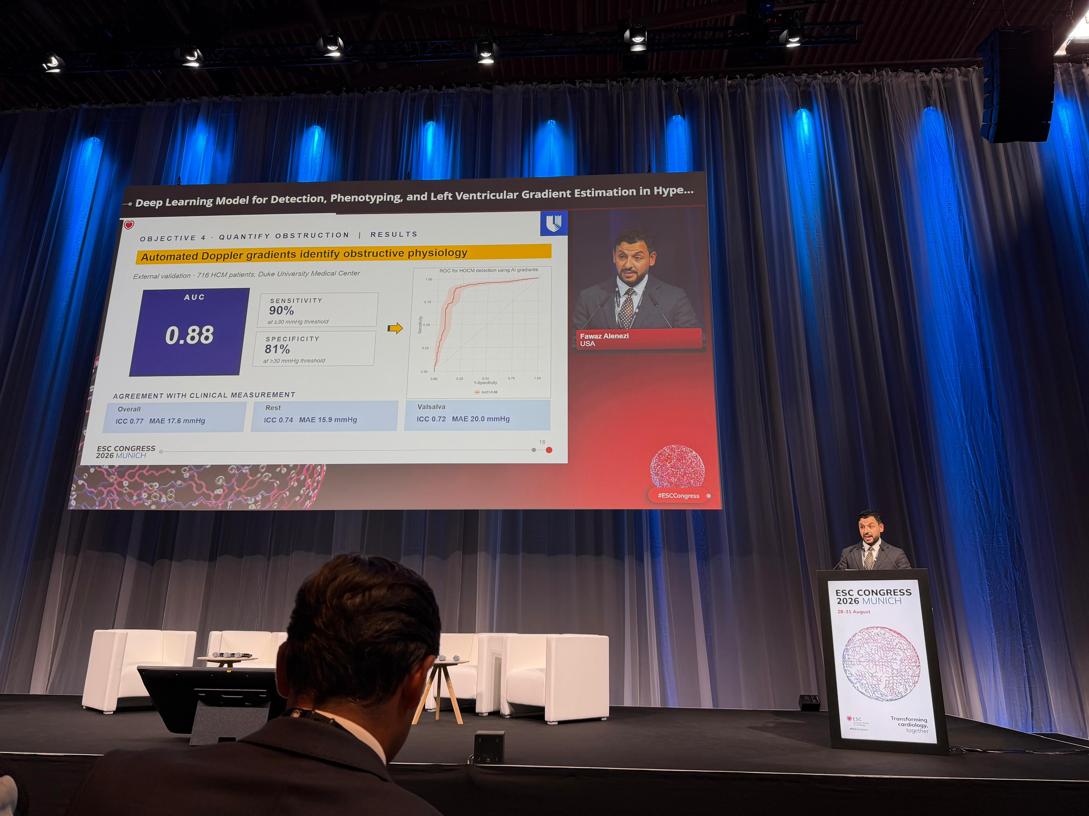
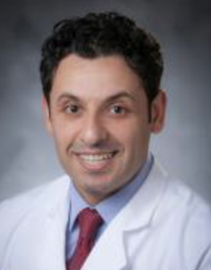
The studies, presented as Late-Breaking Science on Saturday, Aug. 29, at the 2026 European Society of Cardiology Congress in Munich, examined the use of AI with echocardiography to identify and characterize pulmonary hypertension (PH) and hypertrophic cardiomyopathy (HCM). Fawaz Alenezi, MD, MSc, associate professor of medicine in the Division of Cardiology, was first author and presenting investigator for both studies.
Together, the studies involved thousands of patients across multiple institutions, countries, and imaging platforms. Rather than simply using AI to automate measurements already performed by clinicians, the researchers explored whether AI could uncover additional information about disease from echocardiographic images collected during standard care.
In the first study, researchers found that AI could identify pulmonary hypertension — a condition in which blood pressure in the vessels carrying blood from the heart to the lungs is abnormally high — using only a single standard four-chamber echocardiographic video clip. The model did not require Doppler imaging, additional imaging views, or manual measurements.
The model was developed using data from 17,969 Duke patients, including 8,644 patients with PH confirmed through right-heart catheterization, and independently evaluated using patient cohorts from France, Stanford University and China. Its performance increased as disease severity increased, and the model was also able to distinguish among clinically important forms of PH that reflect different underlying pressure patterns in the heart and lung circulation.
The findings suggest that even a single standard echo clip may contain information that can be analyzed in new ways to reveal additional clues about cardiovascular function. In the future, this type of technology could help physicians identify patients who need earlier expert evaluation, including those undergoing echocardiography in community hospitals or other settings where specialized cardiovascular expertise may be less readily available.
The second study focused on hypertrophic cardiomyopathy, a disease in which the heart muscle becomes abnormally thick. The AI model was developed using a multinational cohort of 9,578 patients from Duke and the National Cerebral and Cardiovascular Center in Japan, followed by internal testing in 1,294 patients and independent external validation in 791 patients at Rutgers.
The fully automated system detected HCM from standard echocardiography and distinguished it from other conditions that can cause similar thickening of the heart muscle, including cardiac amyloidosis, aortic stenosis, and hypertensive heart disease. It was also able to recognize different forms of HCM.
A separate AI model evaluated whether the thickened heart muscle was obstructing the flow of blood out of the heart and measured the severity of that obstruction. Together, the models could provide physicians with information not only about whether HCM is present, but also about the form of the disease and how it is affecting heart function.
Taken together, the studies point toward a future in which AI could allow physicians to obtain substantially more clinical information from cardiovascular images already being collected as part of standard care — potentially helping identify disease, determine its type and severity, and guide decisions about further evaluation and care.
“I am especially grateful for the support, mentorship and close collaboration of Sreekanth Vemulapalli and Andrew Wang on the HCM project, and Sudarshan Rajagopal on the pulmonary hypertension project,” Alenezi said. “I am also very grateful to Camille Frazier-Mills and Manesh Patel for their continued support, and to our Duke colleagues and national and international collaborators who made these studies possible.”
Both studies were supported internally by Duke and did not receive external funding.