In 2016, the Duke School of Medicine selected 38 of its faculty for the new Duke Health Scholars and Duke Health Fellows Program. With funds from the Duke University Health System, the program supports the research efforts of early to mid-career clinician-scientists at Duke.
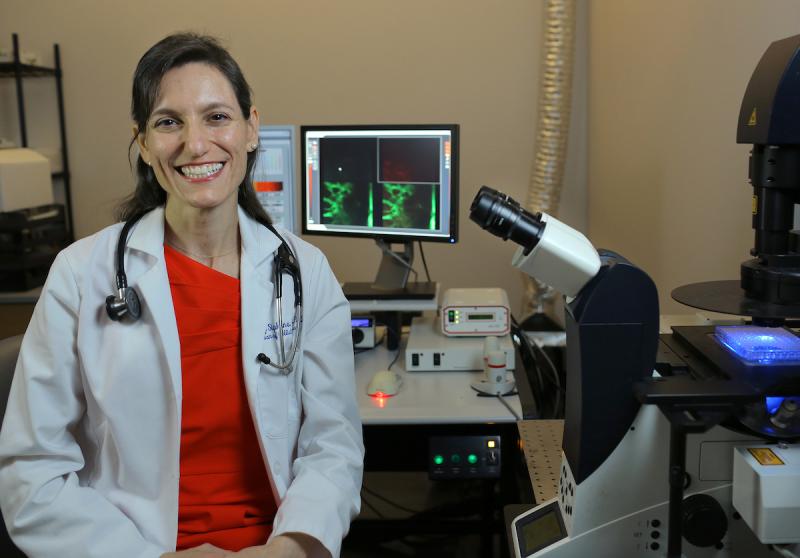
Among the faculty honored are 14 individuals from the Department of Medicine, including Dorothy Sipkins, MD, PhD, associate professor of medicine (Hematologic Malignancies and Cellular Therapy).
Dorothy Sipkins, MD, PhD, is a hematologist-oncologist who studies cancer. But to understand the cleverness of her work, it’s helpful to think of her as an ecologist—a cell ecologist.
Sipkins identifies very specific biological habitats and interactions that allow malignant cells to move, proliferate and survive chemical attacks, traits that too often produce fatal disease.
“I love thinking about what is on the outside of the cell. What the cell is seeing. What the cell is interacting with, the 3-D environment it interacts with,” says Sipkins.
With innovations in imaging and clever experiments, Sipkins is improving our view of what happens within blood vessels and bone marrow that promote relapses of acute leukemia and breast cancer, which could point to new treatments for both.
“By combining genomic, immunohistochemical and functional ex-vivo analyses of xenograft tissues and of primary human samples, she has already made a number of seminal contributions,” said Mary E. Klotman, MD, Dean of Duke’s School of Medicine.
Sipkins has always stepped toward challenges. She decided to become a doctor in elementary school in Carmel, NY, after a teacher’s query about her plans prompted a research trip to the library to explore her options. That goal helped motivate her to work hard enough to become a first-generation college student at Stanford University, where she majored in biology and classics.
Palo Alto was where Sipkins first stepped into a research laboratory. Quickly she liked the analytic and physical demands of the work, whether that required sorting data or inserting catheters into the carotid arteries of hibernating ground squirrels to assess brain ketone metabolism for sleep studies.
Before enrolling in medical school at Stanford, Sipkins moved to Zimbabwe for a year as part of a program that linked Americans with nonprofits needing help. She travelled the country with a social worker providing education and support to the wives of national policemen too often exposed to domestic violence and sexually transmitted disease, including HIV.
Revealing what malignant cells require
Early in Sipkins’ medical studies, an established research chemist in her anatomy class recruited her to help with a project to develop improved contrast media for MRI imaging, an experience that, in time, opened a door for doctoral studies at Stanford.
At that lab bench she invented a molecular-scale flare—a complex of nanoparticle, antibody and MRI contrast media—that migrated to endothelial cells in squamous cell tumor blood vessels in rabbits. Once the complex docked on a targeted receptor, it enhanced tumor visibility on MRI scans.
Sipkins took an improved understanding of both the complexity and predictability of molecular-scale environments to her internal medicine residency at Massachusetts General Hospital. After observing how malignant cells proliferate in patients with relapses of acute leukemia, she became curious about the molecular interactions of leukemic cells.
During a postdoctoral stint, also at Massachusetts General Hospital, Sipkins looked closely at the molecular interactions of leukemic cells in mice, using confocal microscopy to peer through the animal’s paper thin skulls to observe single cells in bone marrow. Leukemic cells, she found, are more likely to nest within bone marrow blood vessels with distinct molecular features, the ability to express an adhesion molecule called E-selectin and the chemoattractant SDF-1 among them.
That suggests that the troublemaking cells depend on micro environments with specific biochemical features.
“The goal of each project is to gain a fundamental understanding of this biologic cross-talk at a level of resolution that defines how, when, and in what context new therapeutic approaches targeting these interactions should be used,” she said.
Sipkins’ laboratory has since found breast cancer cells in mice also favor specific spots in bone marrow, locations that contain the adhesion molecule E-selectin. When her lab dosed the lab mice with plerixafor, a compound that flushes stem cells into the bloodstreams of human bone marrow donors, the breast cancer cells moved into their bloodstream.
That is significant because if that or something like it works in people, that could evict malignant cells from bone marrow, where they can remain dormant for years, and propel them into the circulatory system. There the cancer cells could be more vulnerable to drugs, hormonal therapy an immune attack. “Push them them out into the bloodstream and their Achilles heel may be exposed,” Sipkins said.
The physician scientist is hopeful that clinical trials one day can assess the safety and effectiveness of these strategies and others she hopes to develop.
“Being active across this entire spectrum of bench-to-bedside research is the ideal,” she said.
The series of profiles of our Duke Health Scholars were written by Catherine Clabby, freelance science journalist. Photos are by Ted Richardson.