
What is a gastroenterologist doing studying Parkinson’s Disease?
This is a question that Rodger Liddle, MD, a professor of medicine (Gastroenterology), gets a lot. The answer involves a superhighway, a conversation with a Nobel Laureate, and a plastic foot.
Liddle usually begins the story with the foot.
In his office, carefully situated on a bookshelf among anatomy books, photos from medical school, and colorful knickknacks from international travels, sits the slime-green foot. Approximately five inches long, with an elongated toe bed and a skinny ankle, it almost resembles the snout of a crocodile.
However, to Liddle, it will always look like a foot because it reminds him of the moment that his career took a step in an entirely different direction.
“This is a neuropod,” he explains, holding the plastic 3D-printed model at arm’s length. The strangely shaped cell was discovered by Rashmi Chandra, PhD, and Diego Bohórquez, PhD, in 2011. At the time, Chandra was a long standing member of the Liddle laboratory and Bohórquez was a postdoctoral fellow in his laboratory. They discovered this cell using a confocal microscope to examine the cells that line the gut wall of a mouse.

Rodger Liddle (left) and Diego Bohórquez
For years Liddle has studied how hormones in the gut work. One hormone in particular called cholecystokinin, or CCK, lines the gut wall and is released when cells are jostled around by food. CCK travels through the bloodstream, stimulating digestion and letting the brain know whether the stomach is full and satisfied.
Scientists have long accepted that this process was a conduit for the gut’s connection to the brain. But the cell that the team saw under the microscope that day was different from all the others they’d previously seen hanging out in the gut.
Three years later, in 2014, Bohórquez used electron microscopy to get a 3D image of the cell, and what he saw surprised him. For starters, the cell had neurofilaments, which are typically found in the axons of neurons, or nerve cells – and not normally found in the gut. Secondly, the cell had small clear secretory vesicles, resembling those in neurons that contain neurotransmitters—also not normally found in the gut. On top of that, it was also packed with mitochondria, meaning it required energy. And it had synaptic proteins.
Bohórquez, who trained in the Neurosciences at Duke and the Marine Biological Laboratory, performed other experiments to test this finding. He reached out to colleagues in the Department of Neurobiology including Nicole Calakos, MD, PhD; Marguerita Klein; and Fan Wang, PhD, for guidance.
Wang suggested that he inject rabies virus, a virus that only infects nerve cells, into the gut of a mouse. If the cell in the gut was infected by rabies, it would imply that it was a nerve-like cell. If the rabies virus spread onto an adjacent nerve, it would prove the gut cell was connected by a synapse with the nerve.
Bohórquez injected the virus and found just that - proof that there was a direct circuit—a neurocircuit—between the gut surface and nerves.
Next, he conducted another experiment often called “Two Brains in a Dish” in which a gut cell and a nerve cell were placed into a petri dish together. Within 24 hours, the cells had moved toward one another, touched and then the gut cell withdrew with a connection between them.
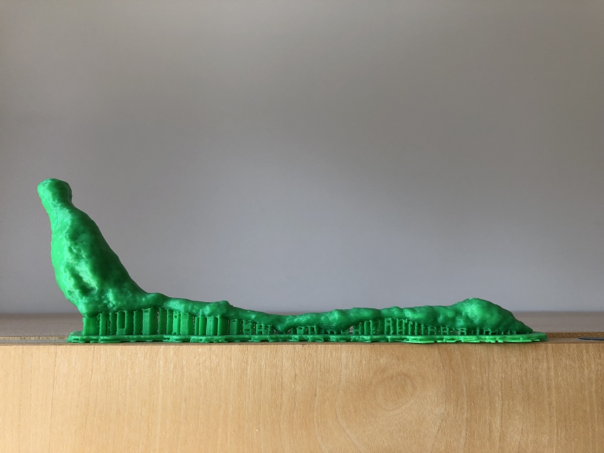
Neuropod model in Dr. Liddle's office
The discovery threw Liddle and Bohórquez for a loop. It suggested a quicker, faster connection between the gut surface and the nervous system, the difference between a superhighway and a slow-moving train.
“We couldn’t contain our excitement,” said Bohórquez. “The term ‘gut feeling’ had always been esoteric, but we were uncovering the basis of a very direct link between the gut and the brain.”
Their discovery was published in the Journal of Clinical Investigation in 2015. From there, the possibilities for new research ventures seemed endless.
Bohórquez, who is now an assistant professor of medicine (Gastroenterology) and neurobiology at Duke, discovered in his laboratory that these cells form a neural circuit conveys signals from nutrients in the gut to the brain in milliseconds. He named these cells neuropod cells. His laboratory studies how the gut senses the caloric value of nutrients to influence the choice of food we eat.
Liddle went another route.
Continue reading on the School of Medicine's Magnify magazine.