At the DCMRC, we are interested in understanding the mechanisms of cardiovascular disease and translating that knowledge into more sensitive and specific cardiovascular imaging.
Current research is aimed towards understanding: 1) the physiologic information portrayed by magnetic resonance images of the heart and 2) the clinical utility of this information. The DCMRC is currently developing additional research projects in collaboration with investigators within Duke University and with investigators at other medical institutions.
Area-at-Risk

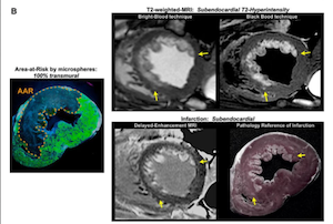
One research area that our group is focused on is imaging the area-at-risk with MRI. The area-at-risk is the myocardium that becomes ischemic after occlusion of its supplying coronary artery in patients suffering myocardial infarction. If area-at-risk could be measured in-vivo, it would allow the direct estimation of myocardial salvage (e.g. the portion of the area-at-risk that survives the ischemic insult), which could then serve as a measure of therapeutic efficacy and as the best surrogate endpoint for randomized trials that test new acute MI therapies. Given the critical importance of measuring the area-at-risk and myocardial salvage, T2-weighted-MRI has been rapidly integrated into clinical trials as a measure of therapeutic efficacy or as a surrogate endpoint.
However, replicating these results proved challenging, and we noted that the pathologic validation for the technique was lacking. Thus, we developed in-vivo models of myocardial infarction in which the area-at-risk could be assessed pathologically. This model allowed the detailed comparison of in-vivo T2-weighted MRI findings with an appropriate gold standard and showed that T2-weighted MRI identifies only the region of acutely infarcted myocardium, rather than the area-at-risk (http://circres.ahajournals.org/content/117/3/254.short).
Testing of novel collagen binding contrast agents
The major goal of this project is the development of a new magnetic resonance imaging probe to obtain high resolution blood flow/perfusion maps without the use of ionizing radiation. We are exploring the utility of an investigational collagen binding molecular imaging probe for detecting myocardial ischemia. The current clinical MR stress perfusion method requires state of the art hardware and ultrafast techniques to image the transit of a bolus of conventional gadolinium contrast media through the myocardium. Thus, image resolution/quality is typically worse than other types of cardiac MR images and the protocol can only be performed with pharmacologically mediated stress. In comparison to conventional MR contrast media, this collagen binding agent has different pharmacokinetics and reversibly binds to collagen in the myocardium in a manner proportional to blood flow and can be detected in the myocardium for minutes to hours. Our group is developing new MR pulse sequences and protocols to take advantage of these properties with the goal that this methodology could be directly translated into use in humans and improve image quality/diagnostic accuracy.
Novel CMR Imaging sequences for improved image quality and diagnostic sensitivity and specificity

One focus of research at the DCMRC is developing new MRI methods for improving the diagnosis of myocardial infarction and other types myocardial damage. The DCMRC developed the current imaging gold-standard for the detection of MI, called Delayed-Enhancement MRI (DE-MRI), which is now used clinically in almost all cardiac magnetic resonance imaging centers worldwide. We have noticed in clinical practice however, that this method has some limitations, and may have reduced sensitivity to small amounts of myocardial injury. We are currently developing a novel technique (FIDDLE) that provides improved visualization of even minimal amounts of injured myocardium. In our initial in-vivo validation studies, we found that this new technique improves the ability to make the diagnosis of MI, and as such will improve the identification and treatment of patients suffering MI and other forms of myocardial damage in clinical practice.
https://www.sciencedirect.com/science/article/pii/S1936878X17309890?via%3Dihub

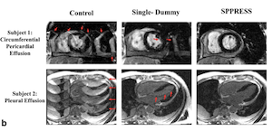
At the DCMRC our research team works closely with the clinical staff to develop new ways to improve CMR image quality. Recently, we have developed methods to reduce ghosting artifacts due to fluids in imaging of myocardial scar. Regions with fluid are commonly encountered in patients referred for cardiac MRI, and include pericardial effusion, pleural effusion, cysts, gastric fluid, and spinal fluid. The artifacts generated by these fluids can overlay important cardiac structures and significantly impair clinical image interpretation. We have developed a new method that effectivelyeliminated these ghosting artifacts allowing for improved image quality. http://onlinelibrary.wiley.com/doi/10.1002/mrm.26554/full

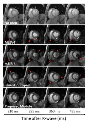
Another recent development by the DCMRC is an improved method for T2-weighting at higher magnetic fields. T2-weighting is a key component of the CMR exam and is used to improve the contrast between the blood and myocardium for coronary angiography. It is also used to detect acute ischemic myocardial injury, and characterize cardiac masses. The technique for T2-weighting used at lower fields results in significant image artifacts at higher fields. Our new module uses a train of adiabatic inversion pulses to provide improved T2-preparation for higher fields. http://onlinelibrary.wiley.com/doi/10.1002/mrm.24564/full